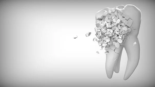
If we cut through an adult human tooth longitudinally from top to bottom, different tissue layers of tooth structure will be evident. Outermost is the enamel followed by dentin. Both of these together form the crown of the tooth that we enjoy observing sparkling in a bright smile! The root portion of the tooth is buried deep into the jaw bone and is covered by the pink gums. The tooth pulp, lying just after dentin, is the heart of our tooth as it harbours blood vessels and nerves as well as provides nutrients to the other tooth tissues. The root of the tooth is covered by a thin tissue layer called cementum. The entire tooth sits inside the bony sockets of our jaw bone and is tightly held in place with the help rope like structures – the periodontal ligaments.

Have you ever wondered how does this intricate arrangement of a human tooth structure is formed inside the human embryo! This very thought gives me goosebumps always! It seems like traversing into the mystic world of finely orchestrated network of cellular and genetic paths and passes filled with starry surprises!
Well, it is fascinating to know that our head and neck are among the most complicated structures that the human embryo forms! This happens through interactions between intermediate structures formed called as pharyngeal arches and all the three embryonic cell layers of endoderm, mesoderm and ectoderm as well as some specialised cells known as the neural crest cells.
If you ever had a chance to closely look into gills of a fish, you may very well imagine an embryonic pharyngeal arch. In the embryo, the pharyngeal arches appear as a series of tissue bands which are externally visible under the anterior portion of the prospective brain

Initially, each pharyngeal arch is formed of identical components which later gives rise to different head and neck structures.
The neural crest cells- are a group of temporary cells that in turn give rise to a variety of other cells which finally differentiate into melanin producing cells (melanocytes) as well as cartilage, bone, smooth muscle of head and neck region. Neural crest cells are unique to vertebrates (animals having backbone) —as are humans.

Our tooth story begins with the aggregation of cells derived from the ectoderm of the first pharyngeal arch and the mesenchyme of neural crest cells (ectomesenchyme). This specialised cellular aggregation, called tooth germ, is the first step in the formation of future teeth. The tooth germ differentiates into three different cellular layers namely — the enamel organ, the dental papilla and the dental sac. Each of these layers gives rise to the different cells and tissues of the future tooth in a series of finely tuned orchestrated events of tooth development which together is referred to as odontogenesis.
We human beings are diphyodont which means we develop two types of teeth in our entire life-time – the set of primary or milk or baby teeth and the permanent set of teeth. Thus, odontogenesis is an on-going process.
You will be surprised to know that all parts of both primary and permanent teeth are formed during the embryonic life itself! It is only that these teeth appear into the mouth at an appropriate time and age after our birth.
Let me make an effort to summarize a brief of our tooth development process:
- Our dentition comprises four groups of teeth each with specialised functions – incisors, canines, premolars and molars.
- The first sign of tooth formation appears as a thickened epithelial band, primary dental lamina, which denotes the tooth rows of the future. Within dental lamina, placodes are formed.
- Placodes are small globular structures which are made of thickened epithelium and neural crest derived mesenchyme. These are considered the first signalling centres of the tooth. Each of the tooth families is formed from each placode.
- As the developmental process proceeds to bud stage, a unique event occurs where the dental epithelium differentiates into two different types of cell groups- the basal cells which remain near the basement membrane peripherally and the other , stellate reticulum, remain centrally located in a loose arrangement. Stellate reticulum is derived from the ectodermal surface cell layers.
- As the primitive tooth bud continues growing, mesenchyme starts accumulating and condensing around it. The dental mesenchyme then differentiates to give rise to dental papilla and dental sac or follicle.
- The cells at the junction of the epithelium and the mesenchyme differentiate into ameloblasts which secrete the tooth enamel.
- The dental papilla gives rise to special cells called odontoblasts which form dentin of the final tooth. The pulp of the tooth arises from the dental papilla being surrounded by the dental epithelium at later stages of development.
- The dental sac housing three groups of specialised cells gives rise to three important structures of the tooth – cementoblasts form the cementum, osteoblasts form the bony sockets (alveolar bone) around the tooth root and fibroblasts form the periodontal tissues.
- The cap and bell stages marks the inception of the regulation of tooth shape and size through special structures called enamel knots. They signal to regulate the growth and cusp shape pattern of the fully formed tooth.

You can well imagine how intricate the entire tooth development process is! The overwhelming information exchange that happens in the background are results of the numerous genetically regulated two-way signalling systems and interactions between the epithelium and the mesenchyme. This specialised group of genetic elements are known as the homeobox genes.

Scientific literatures report the involvement of as many as 300 genes in the process of tooth development!
Most of these genes fall under four major signalling pathways namely –
- BMP / TGFꞵ ( Bone morphogenetic protein / Transforming growth factorꞵ),
- Wnt & Shh
- Fibroblast growth factor
- Eda (Ectodysplasin).
All these pathways integrate with each other in a precise synchrony at different levels.
For example, the function of SHH (Sonic Hedgehog) gene includes signalling tooth initiation and morphogenesis of dental epithelium to ameloblasts (enamel forming cells).
It is imperative that a clearer understanding of such basic biological and genetic mechanisms is of utmost importance. This will empower oral healthcare professionals, researchers and students in diagnosing common dental disorders, analysing rare syndromes, and perhaps in innovating more fruitful solutions to ameliorate the disease burden of the society.
#dentalgenetics #drgargiroygoswami


